
700 Glenhuntly Road
Caulfield VIC 3162
03 9044 4555

700 Glenhuntly Road
Caulfield VIC 3162
03 9044 4555
Peri-Acetabular Osteotomy
A peri-acetabular osteotomy (PAO) is a complex procedure where the hip socket (acetabulum) is reshaped and reorientated into a better position to match up with the position of the ball part of the hip joint (femoral head). This aids to preserve the long-term function of the hip joint and delay the onset of arthritis. The procedure involves cutting of the bone surrounding the acetabulum, reorientating the socket and then holding it in position with wires or screws while the bone heals.
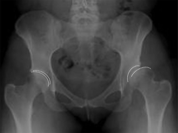
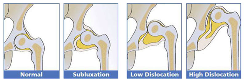
The severity of this abnormal relationship has a fairly wide spectrum. In severe cases there may be such gross abnormalities of the socket and the ball such that the socket is clearly mis-shapen and facing the wrong direction (i.e. backwards or upwards). In these cases, the acetabulum is shaped like a flat, upwardly sloping saucer, compared to the more normal, horizontally-inclined cup shape that usually helps to keep the ball inside the socket. When a patient has a shallow and vertical acetabulum, depending on its degree of severity, it may allow the ball to slide out completely, known as “dislocation” of the hip, or may only allow the ball to slide out partially (thus also remaining partially in contact with the ball too), known as “subluxation”. Clearly, neither of these situations are ideal as they both will often lead to abnormal forces being applied to the joint surfaces, and if left untreated will lead to early hip arthritis and disability. However, if detected before the hip joint cartilage becomes irreversibly damaged, it may be possible to perform a PAO to redirect the socket, permitting the proximal femur to be placed in a more stable position relative to the acetabulum and distributing the forces more normally throughout the redirected hip joint.
Aside from hip dysplasia, the other indication for this procedure is known as acetabular retroversion. In this uncommon condition, the hip socket may be normally shaped, but is facing the wrong direction (usually too far backwards). This has recently been considered a form of femoro-acetabular impingement (FAI), where in some individuals, the parts of the femur and the acetabulum that normally move smoothly without colliding actually bump into each other, and this can result in restriction of movement and may cause damage at the margin of the hip socket to a structure called the acetabular labrum. It is thought that damage to the labrum by this mechanism is one possible cause for a hip arthritis in a young or middle-aged person.
Aside from the routine risks of orthopaedic surgery, there is a small but significant risk of damage to the sciatic nerve which can results in long term loss of sensation in the foot or weakness in the muscles that control the foot, which may result in a limp. There is also small chance that the cut ends of the bone may fail to heal together, called a “non-union”, and a chance that the bone cut may come too close to the cartilage of the hip socket causing damage to it. In some cases, the screws that were implanted to hold the bone fragments may be prominent over a part of the pelvis (near the belt line), and may be required to be removed if they cause significant irritation. This can only be performed once the bone ends have healed, and may not ever be necessary where they are not causing any problems.
Potential Complications of PAO
Aside from the routine risks of orthopaedic surgery, there is a small but significant risk of damage to the sciatic nerve which can results in long term loss of sensation in the foot or weakness in the muscles that control the foot, which may result in a limp. There is also small chance that the cut ends of the bone may fail to heal together, called a “non-union”, and a chance that the bone cut may come too close to the cartilage of the hip socket causing damage to it. In some cases, the screws that were implanted to hold the bone fragments may be prominent over a part of the pelvis (near the belt line), and may be required to be removed if they cause significant irritation. This can only be performed once the bone ends have healed, and may not ever be necessary where they are not causing any problems.
The information above is general. All surgical procedures involve some risk. If you would like advice on your specific condition, please contact Oasis Orthopaedics to make an appointment with one of our specialists.